
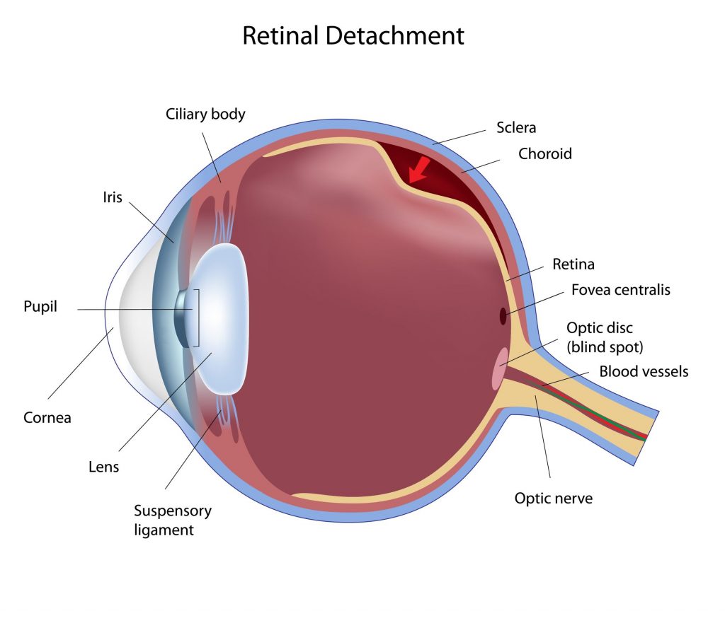
Retinal Detachment is a severe vision-threatening emergency and requires immediate surgical treatment. The retina is the eye’s “camera film” and forms the inside back surface of the eyeball. Please see How the Eyes Work for more details.
Beneath the retina is a layer (the choroid) filled with blood vessels that supply the retina with nourishment. If the retina is torn in one small place, and is treated fairly soon, it will likely regain full function. If a larger retinal area is detached from the blood supply for long enough, its cells will die for lack of oxygen and nutrients. This is why retinal detachment is a medical emergency.
Symptoms of Retinal Detachment
There is no pain, but you would notice changes in your vision:
- Light flashes in one or both eyes
- A darkness or shadow over part of your visual field
- Sudden blurriness
- Sudden appearance of floaters (stringy or spotty shapes that float in your vision and move when you try to focus on them; (they are clumps of cells in the vitreous gel that fills the main part of the eye’s interior)
If you notice any of these changes, contact your eye doctor immediately. Do not wait in hopes that your vision will improve.
How Does Cataract Surgery Lead to Retinal Detachment?
There is a higher risk of retinal detachment after cataract surgery – between five and 16 per 1,000 cataract surgeries, depending on which study you use. In contrast, the risk among those with normal eyes is about five per 100,000.
However, the risk has decreased since ultrasound has been used to break up the lens before removing it. Up until 1990, the lens was removed in one piece, including its capsule. Without the capsule in position, the vitreous gel behind it moved forward. That placed some traction on the retina – the vitreous pulled on it and tore it in some cases, or detached it.
Ultrasound fragments the lens but does not affect its capsule. Your IOL is positioned inside that same capsule and that keeps the vitreous gel in place. Sometimes during cataract surgery, the capsule is broken. That is a potential complication of the surgery. Then the vitreous moves forward and can lead to a retinal tear or detachment.
Cataract Surgery After a Retinal Detachment
If there has been a retinal detachment in the past and that eye has lost some vision, the question is difficult as to whether cataract surgery could then be safely done. Without any cataract surgery, that eye will lose vision completely. But doing the cataract surgery will bring risk of disturbing the vitreous gel and thus endangering the retina again.
Postponing the cataract surgery would probably not be in your best interests, as cataracts are progressive and become larger and harder with time. If you are in this position, your eye surgeon will explain the advantages and disadvantages of going ahead with cataract surgery. Each case is different.
If you would like to consult with a qualified eye surgeon in your area, please contact us as soon as possible and we will supply some names for you.